
Director: Jenny Munson, Ph.D.
Publications
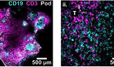
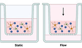
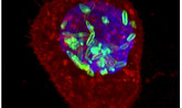
Interstitial fluid flow in an engineered human lymph node stroma model modulates T cell egress and stromal change Open Access
Jennifer H. Hammel, Abhinav Arneja, Jessica Cunningham, Maosen Wang, Sophia Schumaecker, Yamilet Macias Orihuela, Tochukwu Ozulumba, Jonathan M. Zatorski, Thomas J. Braciale, Chance John Luckey, Rebecca R. Pompano, Jennifer M. Munson; Interstitial fluid flow in an engineered human lymph node stroma model modulates T cell egress and stromal change. APL Bioeng. 1 June 2025; 9 (2): 026105. https://doi.org/10.1063/5.0247363
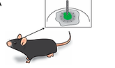
Transient Lymphatic Remodeling Follows Sub-Ablative High-Frequency Irreversible Electroporation Therapy in a 4T1 Murine Model
Esparza, S., Jacobs, E., Hammel, J.H. et al. Transient Lymphatic Remodeling Follows Sub-Ablative High-Frequency Irreversible Electroporation Therapy in a 4T1 Murine Model. Ann Biomed Eng 53, 1148–1164 (2025). https://doi.org/10.1007/s10439-024-03674-y
Interstitial fluid transport dynamics predict glioblastoma invasion and progression
Cora M. Carman-Esparza, Caleb A. Stine, Naciye Atay, Kathryn M. Kingsmore, Maosen Wang, Ryan T. Woodall, Russell C. Rockne, Jessica J. Cunningham, Jennifer M. Munson bioRxiv 2025.03.12.642840; doi: https://doi.org/10.1101/2025.03.12.642840
Ex Vivo Model of Breast Cancer Cell Invasion in Live Lymph Node Tissue
Katerina Morgaenko, Abhinav Arneja, Alexander G. Ball, Audrey M. Putelo, Jennifer M. Munson, Melanie R. Rutkowski, and Rebecca R. Pompano, ACS Pharmacology & Translational Science 2025 8 (3), 690-705, DOI: 10.1021/acsptsci.4c00431
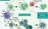
Initiation of primary T cell—B cell interactions and extrafollicular antibody responses in an organized microphysiological model of the human lymph node
Jonathan M. Zatorski, Djuro Raskovic, Abhinav Arneja, Saweetha Kiridena, Tochukwu Ozulumba, Jennifer H. Hammel, Parastoo Anbaei, Jennifer E. Ortiz-Cárdenas, Thomas J. Braciale, Jennifer M. Munson, Chance John Luckey, Rebecca R. Pompano bioRxiv 2025.01.12.632545; doi: https://doi.org/10.1101/2025.01.12.632545
5 - The immune system and its role in the nervous system
Gabriela Geraldo Mendes, Samantha Howerton, Jennifer Munson, 5 - The immune system and its role in the nervous system, Editor(s): Stephanie Willerth, Handbook of Neural Engineering, Academic Press, 2025, Pages 149-177, ISBN 9780323957304, https://doi.org/10.1016/B978-0-323-95730-4.00014-7.
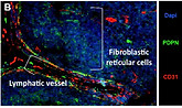
Engineered human lymph node stroma model for examining interstitial fluid flow and T cell egress
Jennifer H Hammel, Abhinav Arneja, Jessica Cunningham, Maosen Wang, Sophia Schumaecker, Yamilet Macias Orihuela, Tochukwu Ozulumba, Jonathan Zatorski, Thomas J Braciale, Chance John Luckey, Rebecca R Pompano, Jennifer M Munson bioRxiv 2024.12.03.622729; doi: https://doi.org/10.1101/2024.12.03.622729

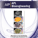
A novel methodology for mapping interstitial fluid dynamics in murine brain tumors using DCE-MRI
Cora Carman-Esparza, Kathryn Kingsmore, Andrea Vaccari, Skylar Davis, Jessica Cunningham, Maosen Wang, Jennifer Munson, A novel methodology for mapping interstitial fluid dynamics in murine brain tumors using DCE-MRI, Methods, Volume 231, 2024, Pages 78-93, ISSN 1046-2023, https://doi.org/10.1016/j.ymeth.2024.09.008.
A Photopolymerizable Hyaluronic Acid-Collagen Model of the Invasive Glioma Microenvironment with Interstitial Flow
Howerton, S., Liang, Y., Hammel, J., Purow, B., Munson, J. A Photopolymerizable Hyaluronic Acid-Collagen Model of the Invasive Glioma Microenvironment with Interstitial Flow. J. Vis. Exp. (212), e66604, doi:10.3791/66604 (2024).
Model discovery approach enables noninvasive measurement of intra-tumoral fluid transport in dynamic MRI Open Access
Ryan T. Woodall, Cora C. Esparza, Margarita Gutova, Maosen Wang, Jessica J. Cunningham, Alexander B. Brummer, Caleb A. Stine, Christine C. Brown, Jennifer M. Munson, Russell C. Rockne; Model discovery approach enables noninvasive measurement of intra-tumoral fluid transport in dynamic MRI. APL Bioeng. 1 June 2024; 8 (2): 026106. https://doi.org/10.1063/5.0190561
Structural and practical identifiability of contrast transport models for DCE-MRI
Conte M, Woodall RT, Gutova M, Chen BT, Shiroishi MS, Brown CE, et al. (2024) Structural and practical identifiability of contrast transport models for DCE-MRI. PLoS Comput Biol 20(5): e1012106. https://doi.org/10.1371/journal.pcbi.1012106
Abstract 2581: Measuring interstitial fluid flow to predict and optimize spatial CAR-T dynamics in the clinic Available
Ryan T. Woodall, Cora Esparza, Christine C. Brown, Jennifer M. Munson, Russell C. Rockne. Measuring interstitial fluid flow to predict and optimize spatial CAR-T dynamics in the clinic [abstract]. In: Proceedings of the American Association for Cancer Research Annual Meeting 2024; Part 1 (Regular Abstracts); 2024 Apr 5-10; San Diego, CA. Philadelphia (PA): AACR; Cancer Res 2024;84(6_Suppl):Abstract nr 2581.
Mitigating reactive oxygen species production and increasing gel porosity improves lymphocyte motility and fibroblast spreading in photocrosslinked gelatin-thiol hydrogels
Tochukwu Ozulumba, Jonathan M. Zatorski, Abhinav Arneja, Jennifer H. Hammel, Thomas J. Braciale, Chance J. Luckey, Jennifer M. Munson, Rebecca R. Pompano
bioRxiv 2024.01.14.574282; doi: https://doi.org/10.1101/2024.01.14.574282
Demonstration of chemotherapeutic mediated lymphatic changes in meningeal lymphatics in vitro, ex vivo, and in vivo
L. Monet Roberts, Jennifer H Hammel, Francesca Azar, Tzu-Yu (Alkaid) Feng, Jessica J. Cunningham, Melanie Rutkowski, Jennifer Munson
bioRxiv 2024.01.06.574460; doi: https://doi.org/10.1101/2024.01.06.574460
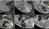
Pregnancy-induced remodeling of the murine reproductive tract: a longitudinal in vivo magnetic resonance imaging study
Suarez, A.C., Gimenez, C.J., Russell, S.R. et al. Pregnancy-induced remodeling of the murine reproductive tract: a longitudinal in vivo magnetic resonance imaging study. Sci Rep 14, 586 (2024). https://doi.org/10.1038/s41598-023-50437-1
Structural and practical identifiability 1 of contrast transport models for DCE-MRI
Martina Conte, Ryan T. Woodall, Margarita Gutova, Bihong T. Chen, Mark S. Shiroishi, Christine E. Brown, Jennifer M. Munson, and Russell C. Rockne, bioRxiv, Page v282, https://doi.org/10.1101/2023.12.19.572294
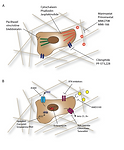
TMIC-21. SPHINGOSINE-1-PHOSPHATE RECEPTOR 3 (S1PR3) IN THE GLIOBLASTOMA TUMOR MICROENVIRONMENT MAY PLAY A ROLE IN IMMUNOSUPPRESSION
Sharon Michelhaugh, Jennifer Munson, TMIC-21. SPHINGOSINE-1-PHOSPHATE RECEPTOR 3 (S1PR3) IN THE GLIOBLASTOMA TUMOR MICROENVIRONMENT MAY PLAY A ROLE IN IMMUNOSUPPRESSION, Neuro-Oncology, Volume 25, Issue Supplement_5, November 2023, Page v282, https://doi.org/10.1093/neuonc/noad179.1087
TMIC-64. CHARACTERIZATION OF THE TUMOR MICROENVIRONMENT IN DIFFUSE MIDLINE GLIOMAS (DMG) FOR 3D IN VITRO MODEL BUILDING
Sarah Kremer, Rhea John, Peng Jin, Sharon Michelhaugh, Jessica Cunningham, Samantha Howerton, Augustine Eze, Javad Nazarian, Jennifer Munson, TMIC-64. CHARACTERIZATION OF THE TUMOR MICROENVIRONMENT IN DIFFUSE MIDLINE GLIOMAS (DMG) FOR 3D IN VITRO MODEL BUILDING, Neuro-Oncology, Volume 25, Issue Supplement_5, November 2023, Pages v292–v293, https://doi.org/10.1093/neuonc/noad179.1130
580 Mapping interstitial fluid flow in the brain to improve CAR T cell trafficking and efficacy
Margarita Gutova, Ryan Woodall, Eric Ma, Cora Esparza, Vanessa Salvary, Brenda Aguilar, Renate Starr, Behnam Badie, Darya Alizadeh, Jennifer M Munson, Russell C Rockne, Christine Brown - 580 Mapping interstitial fluid flow in the brain to improve CAR T cell trafficking and efficacy: Journal for ImmunoTherapy of Cancer 2023;11:.
Model discovery approach enables non-invasive measurement of intra-tumoral fluid transport in dynamic MRI
Ryan T. Woodall, Cora C. Esparza, Margarita Gutova, Maosen Wang, Jessica J. Cunningham, Alexander B. Brummer, Caleb A. Stine, Christine C. Brown, Jennifer M. Munson, Russell C. Rockne
bioRxiv 2023.08.28.554919; doi: https://doi.org/10.1101/2023.08.28.554919
Development of a Synthetic, Injectable Hydrogel to Capture Residual Glioblastoma and Glioblastoma Stem-Like Cells with CXCL12-Mediated Chemotaxis
Z. M. Khan, J. M. Munson, T. E. Long, E. Vlaisavljevich, S. S. Verbridge, Development of a Synthetic, Injectable Hydrogel to Capture Residual Glioblastoma and Glioblastoma Stem-Like Cells with CXCL12-Mediated Chemotaxis. Adv. Healthcare Mater. 2023, 12, 2300671. https://doi.org/10.1002/adhm.202300671
Modeling lymphangiogenesis: Pairing in vitro and in vivo metrics
Suarez AC, Hammel JH, Munson JM. Modeling lymphangiogenesis: Pairing in vitro and in vivo metrics. Microcirculation. 2023; 30:e12802. doi:10.1111/micc.12802
Non-invasive measurement of intra-tumoral fluid dynamics with localized convolutional function regression
Ryan T. Woodall, Cora C. Esparza, Margarita Gutova, Maosen Wang, Jessica J. Cunningham, Alexander B. Brummer, Caleb A. Stine, Christine C. Brown, Jennifer M. Munson, Russell C. Rockne
bioRxiv 2023.08.28.554919; doi: https://doi.org/10.1101/2023.08.28.554919
Fluids and flows in brain cancer and neurological disorders
Jin, P., & Munson, J. M. (2023). Fluids and flows in brain cancer and neurological disorders. WIREs Mechanisms of Disease, 15(1), e1582. https://doi.org/10.1002/wsbm.1582
Towards spatially-organized organs-on-chip: Photopatterning cell-laden thiol-ene and methacryloyl hydrogels in a microfluidic device
Jennifer E. Ortiz-Cárdenas, Jonathan M. Zatorski, Abhinav Arneja, Alyssa N. Montalbine, Jennifer M. Munson, Chance John Luckey, Rebecca R. Pompano, Towards spatially-organized organs-on-chip: Photopatterning cell-laden thiol-ene and methacryloyl hydrogels in a microfluidic device, Organs-on-a-Chip, Volume 4, 2022, 100018, ISSN 2666-1020, https://doi.org/10.1016/j.ooc.2022.100018.
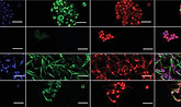
Fusobacterium nucleatum induces proliferation and migration in pancreatic cancer cells through host autocrine and paracrine signaling
Barath Udayasuryan, Raffae N Ahmad, Tam TD Nguyen, Ariana Umaña, LaDeidra Monét Roberts, Polina Sobol, Stephen D Jones, Jennifer M Munson, Daniel J Slade, Scott S Verbridge Fusobacterium nucleatum induces proliferation and migration in pancreatic cancer cells through host autocrine and paracrine signaling.Sci. Signal.15,eabn4948(2022).DOI:10.1126/scisignal.abn4948

Patient-designed models of the invasive edge of glioblastoma
RC Cornelison, JX Yuan, KM Tate, A Petrosky†, GF Beeghly, M Bloomfield, SC Schwager†, AL Berr†, D Cimini, FF Bafakih, JW Mandell, BW Purow, BJ Horton, JM Munson*. A patient-designed tissue-engineered model of the infiltrative glioblastoma microenvironment, NPJ Precision Oncology.6(54) 2022. Available: https://rdcu.be/cXtxU
.png)
Cell migration in response to flow and endothelial cell interactions
L. M. Roberts, M. J. Perez, K. N. Balogh, G. Mingledorff, J. V. Cross, and J. M. Munson, “Myeloid Derived Suppressor Cells Migrate in Response to Flow and Lymphatic Endothelial Cell Interaction in the Breast Tumor Microenvironment,” Cancers, vol. 14, no. 12, Art. no. 12, Jan. 2022, doi: 10.3390/cancers14123008.
.png)
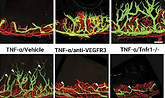
Lymphangiogenesis to be prevented with Anti-VEGFR3 therapy
A. R. Harris et al., “Platinum Chemotherapy Induces Lymphangiogenesis in Cancerous and Healthy Tissues That Can be Prevented With Adjuvant Anti-VEGFR3 Therapy,” Frontiers in Oncology, vol. 12, 2022, Accessed: Jun. 27, 2022. [Online]. Available: https://www.frontiersin.org/article/10.3389/fonc.2022.801764
_edited_edited.jpg)
Repeatability of tumor perfusion kinetics
R. T. Woodall et al., “Repeatability of tumor perfusion kinetics from dynamic contrast-enhanced MRI in glioblastoma,” Neuro-Oncology Advances, vol. 3, no. 1, p. vdab174, Jan. 2021, doi: 10.1093/noajnl/vdab174.

Gradient formation in interstitial fluid flow environments
C. A. Stine and J. M. Munson, “Autologous Gradient Formation under Differential Interstitial Fluid Flow Environments,” Biophysica, vol. 2, no. 1, Art. no. 1, Mar. 2022, doi: 10.3390/biophysica2010003.

Tissue engineering models of breast cancer
J. A. McGuire et al., “Tear propagation in vaginal tissue under inflation,” Acta Biomaterialia, vol. 127, pp. 193–G. F. Beeghly, C. Thomas, J. X. Yuan, A. R. Harris, and J. M. Munson, “Designing Patient-Driven, Tissue-Engineered Models of Primary and Metastatic Breast Cancer,” Bioengineering, vol. 9, no. 2, Art. no. 2, Feb. 2022, doi: 10.3390/bioengineering9020044.

Engineering advances for women’s health
R. De Vita and J. Munson, “Special Issue on the Advances in Engineering for Women’s Health,” Ann Biomed Eng, vol. 49, no. 8, pp. 1785–1787, Aug. 2021, doi: 10.1007/s10439-021-02837-5.

Tear propagation in vaginal tissue
J. A. McGuire et al., “Tear propagation in vaginal tissue under inflation,” Acta Biomaterialia, vol. 127, pp. 193–204, Jun. 2021, doi: 10.1016/j.actbio.2021.03.065.

Ultrasound promotes nanoparticle dispersion and transfection
Curley T., Colleen et al. Augmentation of brain tumor interstitial flow via focused ultrasound promotes brain-penetrating nanoparticle dispersion and transfection. Science Advances, vol. 6, no. 18, p. eaay1344, May 2020, doi: 10.1126/sciadv.aay1344.

Measuring interstitial fluid flow in glioblastoma patients
Atay, Chatterjee, et al. Utilizing Dynamic Contrast-Enhanced Magnetic Resonance Imaging (DCE-MRI) to Analyze Interstitial Fluid Flow and Transport in Glioblastoma and the Surrounding Parenchyma in Human Patients. Pharmaceutics. 13 (2): 212 (2021). doi: 10.3390/pharmaceutics13020212

Focused Ultrasound-mediated blood brain barrier opening increases interstitial fluid flow
Curley, et al. Augmentation of brain tumor interstitial flow via focused ultrasound promotes brain-penetrating nanoparticle dispersion and transfection. Science Advances.6(18): eaay1344 (2020). DOI: 10.1126/sciadv.aay1344

Review: Engineering the tumor microenvironment
LM Roberts, JM Munson, Modulating microenvironments for treating glioblastoma. Current tissue microenvironment reports. 1, 99-111 (2020)

Review: Studying fluid flow in the brain
KC Chatterjee, CA Carman-Esparza, JM Munson, Methods to measure, model, and manipulate fluid flow in the brain. Neuroscience Methods. 333: 108541. (2020) https://doi.org/10.1016/j.jneumeth.2019.108541

Review: Linking CED to interstitial flow
CA Stine, JM Munson, Convection enhanced delivery: role of increased interstitial fluid flow. Frontiers: Oncology. 9: 966 (2019).


Review: Systems for testing drugs for nervous disorders
1. KM Tate, JM Munson, Assessing drug response in engineered neural microenvironments. Brain Research Bulletin. 150: 21-34 (2019). DOI: 10.1016/j.brainresbull.2019.04.027

Increased fluid flow in vivo leads to increased glioma invasion
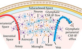
RC Cornelison, KM Kingsmore, CR Brennan, JM Munson, Functional aspects of meningeal lymphatics in ageing and Alzheimer’s disease. Nature. (2018). DOI: 10.1038/s41586-018-0368-8.

Increasing meningeal lymphangiogenesis to boost memory
S Da Mesquita, A Louveau, A Vaccari, I Smirnov, RC Cornelison, KM Kingsmore, C Contarino, S Onengut-Gumuscu, E Farber, D Raper, KE Viar, W Baker, N Dabhi, G Oliver, S Rich, JM Munson, CC Overall, ST Acton, J Kipnis, Functional aspects of meningeal lymphatics in ageing and Alzheimer’s disease. Nature. (2018). DOI: 10.1038/s41586-018-0368-8.
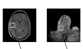
Mapping interstitial flow in glioblastoma with MRI
Kathryn M. Kingsmore, Andrea Vaccari, Daniel Abler, Sophia X. Cui, Frederick H. Epstein, Russell C. Rockne, Scott T. Acton, and Jennifer M. Munson, MRI analysis to map interstitial flow in the brain tumor microenvironment. (2018) APL Bioengineering 18(1), 718. DOI: 10.1063/1.5023503

Common chemotherapy promotes microenvironmental changes
Alexandra R Harris, Matthew J Perez, Jennifer M. Munson , Docetaxel facilitates lymphatic-tumor crosstalk to promote lymphangiogenesis and cancer progression. (2018) BMC Cancer 18(1), 718. DOI: 10.1186/s12885-018-4619-8

How can we use in vitro models to inform biomaterial use in glioma?
R Chase Cornelison, Jennifer M. Munson, Perspective on Translating Biomaterials Into Glioma Therapy: Lessons From in Vitro Models. (2018) Frontiers Materials 5, 27. DOI: 10.3389/fmats.2018.00027

Methods for tissue engineering cancer in vitro
Alexandra R Harris, Jessica X. Yuan, Jennifer M. Munson , Assessing multiparametric drug response in tissue engineered tumor microenvironment models, Methods (2017) 134-135: 20-31. DOI: 10.1016/j.ymeth.2017.12.010

Breast cancer chemotherapy response varies at the border
Daniel K. Logsdon, Garrett F. Beeghly, Jennifer M. Munson, Chemoprotection Across the Tumor Border: Cancer Cell Response to Doxorubicin Depends on Stromal Fibroblast Ratios and Interstitial Therapeutic Transport. Cellular and Molecular Bioengineering (2017) 10(5): 463-481. DOI: 10.1007/s12195-017-0498-3

Quantitative analysis of the glioma microenvironment
Jessica X. Yuan, Jennifer M. Munson, Quantitative Immunohistochemistry of the Cellular Microenvironment in Patient Glioblastoma Resections. Journal of Visualized Experiments (2017). DOI: 10.3791/56025

The cellular microenvironment of GBM can predict survival
Jessica X. Yuan BS, Fahad F. Bafakih MD, James W. Mandell MD, PhD, Bethany J. Horton PhD, Jennifer M. Munson PhD, Quantitative Analysis of the Cellular Microenvironment of Glioblastoma to Develop Predictive Statistical Models of Overall Survival. Journal of Neuropathology and Experimental Neurology. (2016) 75(12): 1110-1123. DOI: 10.1093/jnen/nlw090

Multiple mechanisms underlie interstitial flow-stimulated invasion
KM Kingsmore, DK Logsdon, DJ Floyd, BW Purow, JM Munson, Interstitial flow differentially increases patient-derived glioblastoma stem cell invasion via CXCR4, CXCL12, and CD44-mediated mechanisms. Integrative Biology (2016) 8: 1246-1260 . DOI: 10.1039/C6IB00167J

Review: Macrophages' role in angiogenesis and lymphangiogenesis
BA Corliss, MS Azimi, JM Munson, SM Peirce, WL Murfee, Macrophages: an inflammatory link between angiogenesis and lymphangiogenesis. Microcirculation (2016) 23(2): 95-121. DOI: 10.1039/C6IB00167J

The role of interstitial flow in cancer progression
JM Munson, AC Shieh, Interstitial fluid flow in cancer: implications for disease progression and treatment. Cancer Management and Research 6: 317-328 (2014). DOI: 10.2147/CMAR.S65444
Pre-2014
New therapeutics against glioma invasion
JM Munson, MY Bonner, L Fried, JL Arbiser, RV Bellamkonda, Identifying new small molecule anti-invasive compounds for glioma treatment. Cell Cycle 12 (14):1-10 (2013).
Interstitial flow enhances glioma invasion
JM Munson, RV Bellamkonda & MA Swartz, Interstitial flow increases glioma invasion via CXCR4-dependent autologous chemotaxis in a 3D microenvironment. Cancer Research 73(5): 1536-1546 (2013)

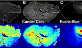
Using nanoparticles for intraoperative glioma ID
BR Roller, JM Munson, PA Santangelo, B Brahma, RV Bellamkonda, Evans blue nanocarriers visually demarcate margins of invasive gliomas, Drug delivery and Translational Research (2013).

New nanoparticle therapy stops glioma invasion
JM Munson, L Fried, SA Rowson, MY Bonner, L Karumbaiah, B Diaz, SA Courtneidge, UG Knaus, DJ Brat, JL Arbiser, RV Bellamkonda, Anti-invasive adjuvant therapy with Imipramine Blue enhances chemotherapeutic efficacy against glioma. Science Translational Medicine 4, 127ra36 (2012).